
AI cell imaging made faster
Encore is an imaging and analysis platform for laboratories that run a range of assays requiring high capacity, intelligent automation, and flexible workflows on a single compact system.


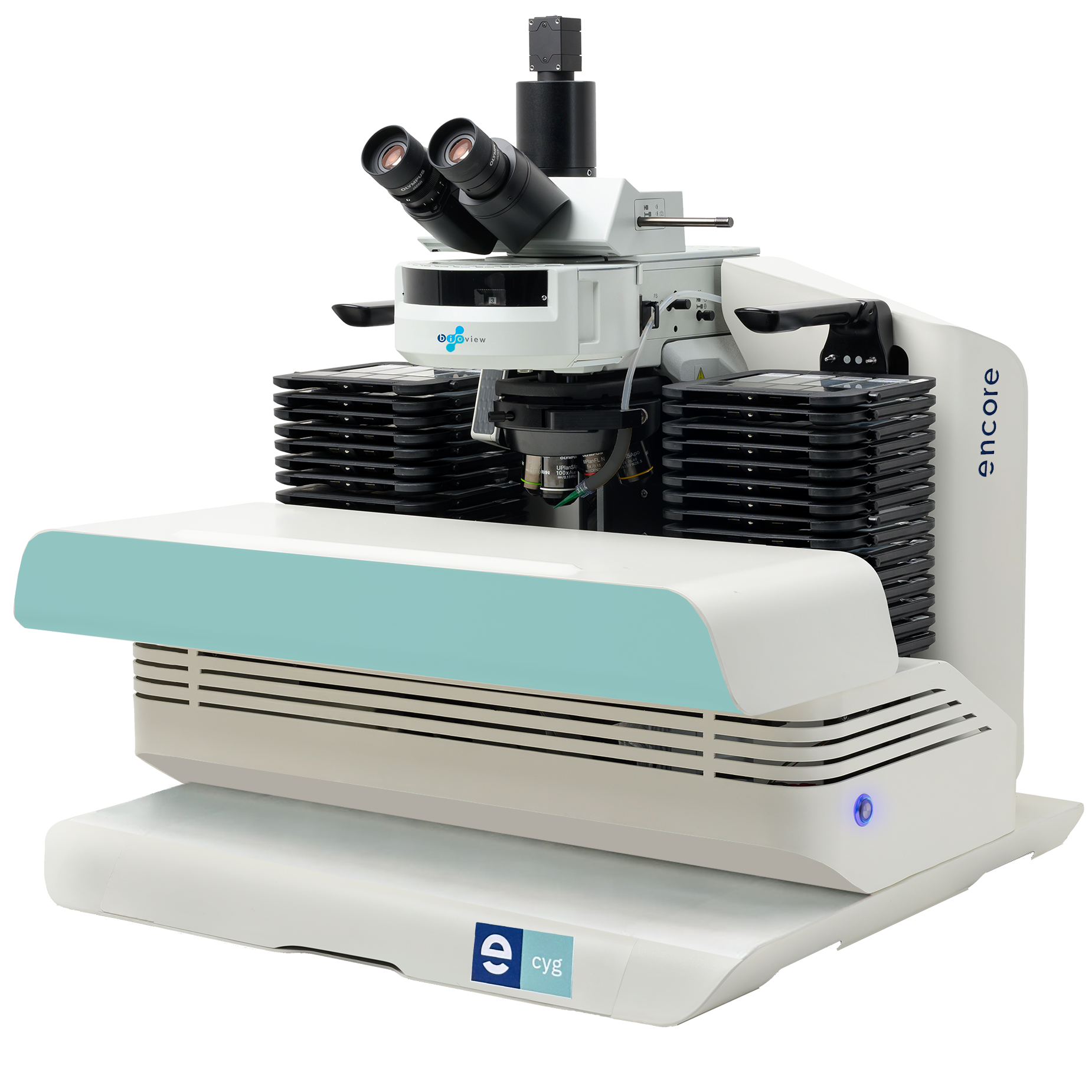
Everything a modern lab
needs - in one platform.
Exceptional imaging
Capture precise, consistent images across fluorescence and brightfield applications with high-resolution optics, stable illumination, and automated imaging parameters.
Intelligent automation
Encore recognizes each test, applies the appropriate protocol, switches between imaging modes, and analyzes images as they are captured.
Higher throughput
Keep cases moving with 60- or 120-slide capacity and unattended operation, reducing hands-on time and supporting continuous scanning day and night.
Review from anywhere
Review, analyze, and sign off on cases locally or remotely through a standard web browser, with no software or plug-ins required.
One batch.
Multiple applications.
Encore brings different applications and imaging modes, together in one fast workflow.
- 01
Load a mixed batch
Prepare slides in the wet lab using the robust detachable magnetic cassette, then combine applications into a single uninterrupted batch.
- 02
Encore verifies every slide
A dual-modality identification system reads the barcode scan definitions while capturing a visual record of the label printed on the slide.
- 03
Imaging adapts automatically
Encore switches the protocol for each application.
- 04
Cases become ready for review
Images are processed as they are captured, allowing teams to begin analysis while Encore continues scanning.
Built around
your test menu
Encore brings karyotyping, FISH, cytology, pathology, and rare-cell applications together on one automated imaging platform.
Cytogenetics
Automated imaging for karyotyping and FISH workflows across cytogenetic samples.
Not sure which applications fit your workflow?
Precision engineered
into every scan.
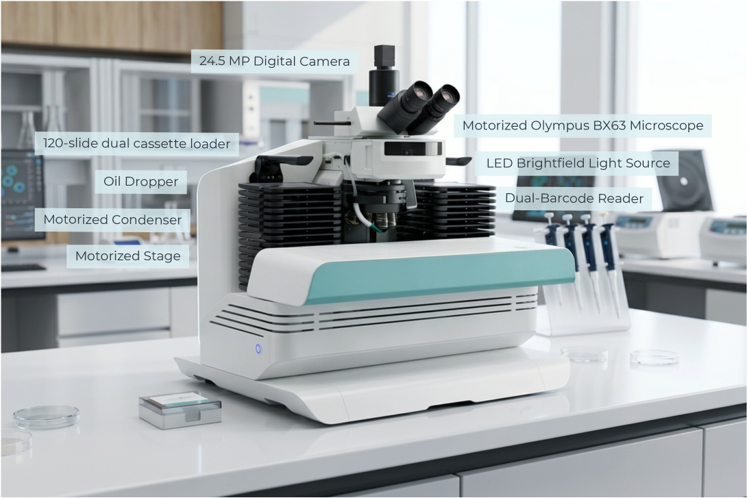
Every component works together to support accurate and consistent optical performance.
Dual-Modality Barcode Verification System
Enhanced sample traceability and error prevention through a dual-modality barcode identification mechanism, combining:
- •Barcode Reader — for fast and accurate digital extraction of the case ID and scan definitions
- •Imaging Camera — for capturing a visual record of the barcode physically printed on the slide
LED Fluorescent Light Source
- •High-power LED fluorescent light source
- •Consistently produces outputs of ~70% of initial, out-of-the-box optical power for more than a decade of use
- •Vibration and heat-free light delivery via liquid light guide
- •Alignment-free
High Resolution 24.5MP Digital Camera
- •CMOS sensor offers a resolution of 5320 × 4600 pixels, enabling detailed visualization of chromosomal structures and fluorescent signals
- •High dynamic capture of both bright and dim signals within the same image, crucial for samples with varying fluorescence intensities
Olympus UPLXAPO Oil Immersion Objectives for High-Resolution Imaging
- •Enhancing signal-to-noise ratio in low-light fluorescence imaging
- •Advanced apochromatic correction across a wide spectral range, including near-UV and far-red
Designed to work with
your laboratory.
Integration and deployment options can be configured around your laboratory environment.
Configure Encore
around your laboratory.