

How AI Supports Automated Cellular Imaging: Detection, Segmentation and Classification
Detection, segmentation and classification are the three tasks behind most AI-assisted cell imaging. What each one does, what training data they need, and where expert review stays essential.
When artificial intelligence is described as part of an automated imaging system, the phrase covers several distinct computational tasks. In cellular imaging, most of them fall into three families: finding objects, outlining them precisely, and assigning them to a category. Understanding which task a tool performs makes it far easier to judge what it can reasonably contribute to a laboratory workflow.
None of these tasks replaces interpretation. They prepare structured material, located objects, measured boundaries, proposed labels, that a qualified professional then reviews, corrects and signs out under the laboratory's own procedures.
Detection: locating objects in the image
Detection, sometimes called localization, answers a simple question: where in this image are the objects of interest? The output is typically a set of coordinates or bounding regions marking candidate nuclei, cells, metaphase spreads or fluorescent signals. Detection does not describe an object in detail; it establishes that something worth examining is present at a given position.
In a scanning workflow this is what turns a large slide area into a manageable list of candidates. Instead of navigating the whole slide, the analyst begins from positions the system has already flagged, and can dismiss unsuitable candidates quickly.
Segmentation: where the boundaries are
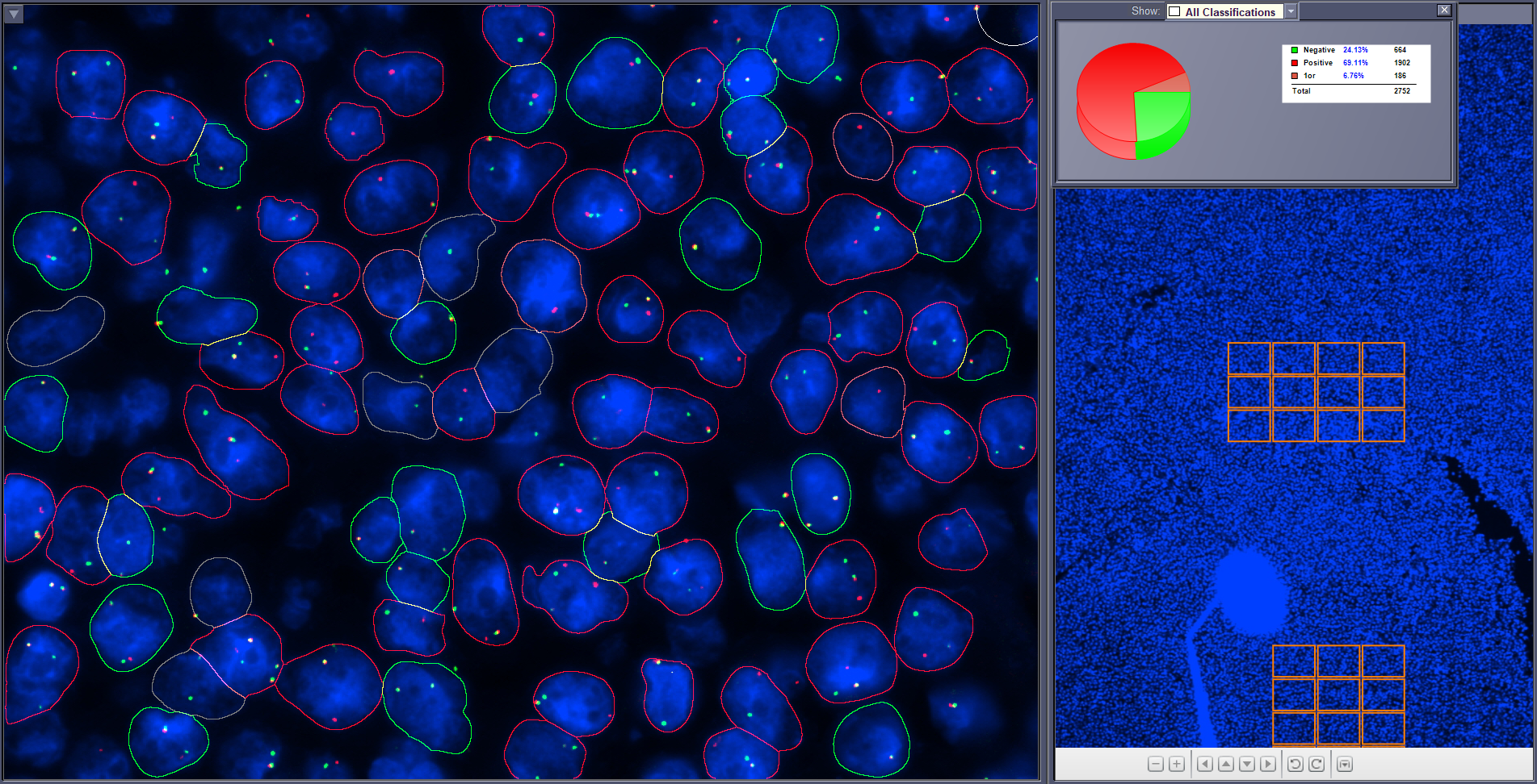
Segmentation goes further than detection by assigning image pixels to specific objects. Rather than a box around a nucleus, segmentation produces its outline. That distinction matters whenever a measurement depends on the boundary: nuclear area, shape descriptors, signal-to-nucleus assignment, or the separation of touching and overlapping objects.
Because downstream numbers inherit the boundary, segmentation errors propagate. Two nuclei merged into one changes counts; a boundary drawn too tightly changes area and intensity statistics. This is one reason reviewable overlays are valuable, an analyst can see the outline the system used, not only the number it produced.
Classification: assigning objects to groups
Classification places a detected or segmented object into one of a set of predefined groups , for example, sorting chromosomes into their expected classes, or separating nuclei from debris and artefacts. A classifier can only use the categories it was trained on; anything outside that set has to be handled by the reviewer or by an explicit rejection category.
What makes training data difficult in cell imaging
Models learn from examples, and biological images present several forms of variation that examples must cover if the model is to behave predictably in routine use.
- Viewpoint and alignment: objects appear at arbitrary rotations and positions, and chromosomes in particular may be bent, straight, or foreshortened.
- Natural deformation and biological variability: cells and chromosomes are not rigid shapes, and normal morphology spans a wide range.
- Occlusion and overlap: touching nuclei and crossing chromosomes hide part of the object that a model needs to see.
- Inter-class and intra-class variation: some categories look similar to each other, while members of the same category can differ noticeably between preparations, stains and sites.
Practically, this means data that reflects the laboratory's own preparations, stains and imaging conditions is more informative than a larger but less representative dataset.
A concrete example: karyotyping
Karyotyping uses all three tasks in sequence. Detection locates metaphase spreads on the slide and chromosomes within a spread. Segmentation separates individual chromosomes, including touching or overlapping ones. Classification proposes a class for each chromosome so that a karyogram layout can be presented.
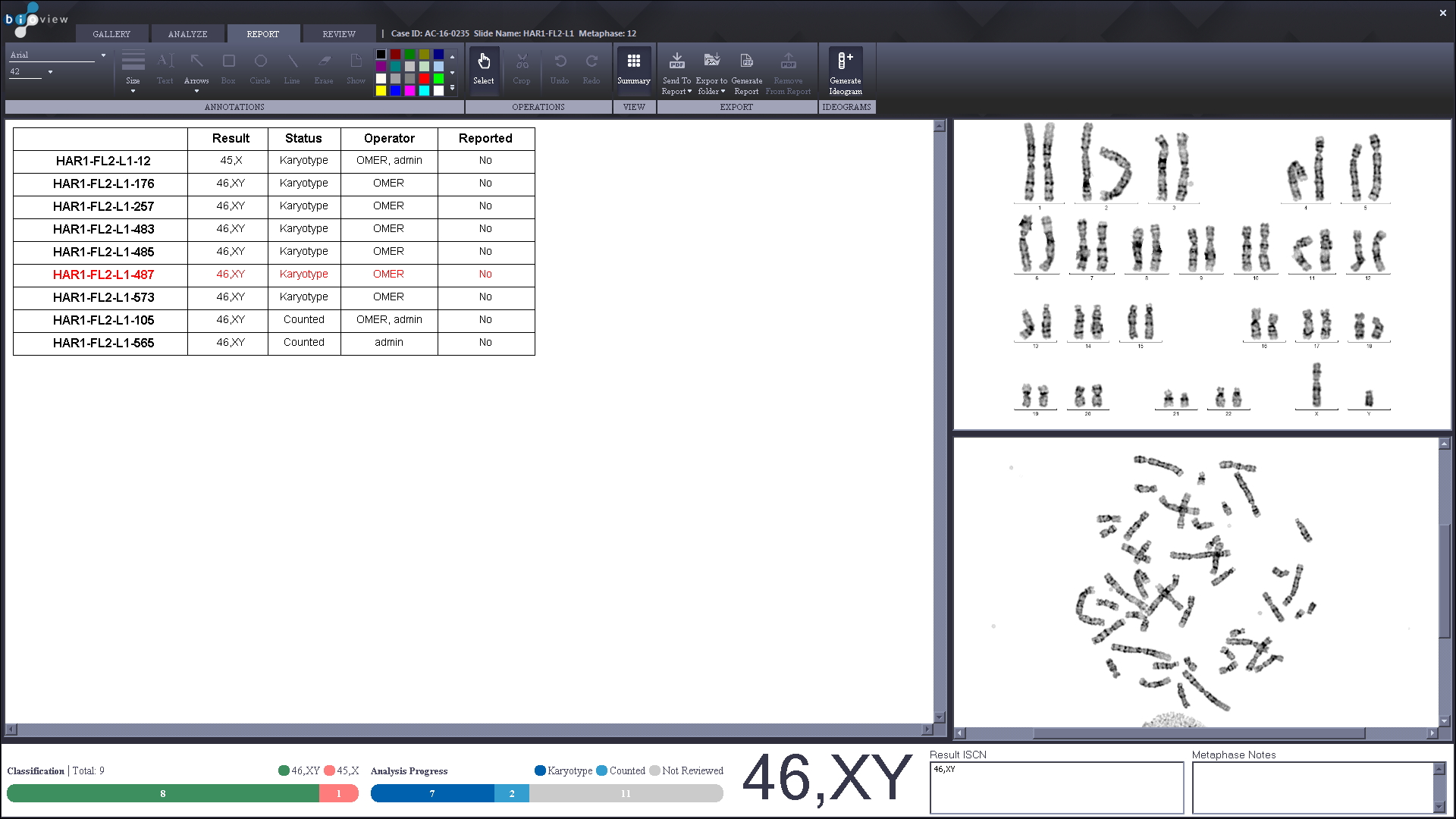
What the analyst receives is a proposal. Reassigning a chromosome, correcting a split, or rejecting a spread entirely are all normal parts of the workflow, and the final karyotype remains the professional's determination.
Reported accuracy in routine use
Accuracy figures are only meaningful with their scope attached. In a cytogenetics laboratory using BioView for karyotype analysis of bone marrow and peripheral blood cultures, the laboratory director reported automated analysis accuracy close to 99.9%, explicitly qualified as subject to sample quality, and with every cell still reviewed by a specialist before sign-out.
Analysis with an accuracy rate close to 99.9% (subject to sample quality). Every cell is still reviewed by a specialist, but the process is fast.
That figure describes one laboratory's experience with its own sample types, preparations and procedures. It is not a general performance specification, and it does not extend to other assays, sample types or laboratories. Any laboratory adopting automated analysis should establish its own performance against its own material.
What laboratories should consider
- Imaging consistency: stable illumination, focus and exposure settings give a model the conditions it was trained for.
- Representative training and evaluation data covering the sample types, stains and preparation quality the laboratory actually handles.
- Workflow validation against the laboratory's own material and procedures before routine use.
- Human review at defined points, with the ability to inspect and correct what the system proposed.
- Ongoing monitoring, so that changes in reagents, protocols or instruments are noticed rather than absorbed silently.
Framed this way, AI in cellular imaging is a workflow technology. It changes how much manual navigation and preparation a case requires, and how consistently material is presented for review while the analytical decision, and the responsibility for it, stay with the laboratory.