

How AI “Sees” a Cell: The Image Features Behind Automated Analysis
An algorithm does not look at a cell the way a pathologist does. It measures features. Understanding which features drive automated analysis explains both its usefulness and its limits.
It is tempting to describe an AI model as “looking at” a cell. The metaphor is convenient, but it hides an important difference. A pathologist or cytogeneticist reads an image in context: the clinical question, the preparation, the rest of the slide, and years of comparable cases. An algorithm works from measurable properties of pixels.
Knowing which properties are in play makes automated output much easier to judge, and much easier to review productively.
The features behind automated analysis
Most cell-image models operate, directly or indirectly, on a familiar set of measurable characteristics.
- Nuclear size: area or diameter derived from the segmented boundary.
- Shape: circularity, elongation and boundary regularity.
- Texture: the pattern of intensity variation within an object, which reflects chromatin appearance and staining.
- Intensity: brightness in each acquired channel, and its distribution within the object.
- Signal distribution: how discrete fluorescent signals are positioned and spaced inside a nucleus.
- Spatial relationships: proximity between objects, clustering, and whether nuclei touch or overlap.
Modern models may learn their own internal representations rather than using a hand-written list, but the information available to them still comes from these same physical properties of the image.
Contextual interpretation versus feature analysis
A professional can recognise that an unusual appearance is explained by a fixation artefact, an unexpected sample type, or a known feature of the case. A feature-based system has no access to that context. It reports what the pixels support, within the categories it was trained to distinguish.
That difference is not a flaw so much as a boundary. Algorithmic analysis is consistent and tireless across thousands of objects; contextual judgement is what determines whether a consistent measurement is also a meaningful one.
Why input quality shapes the output
Because everything downstream derives from the image and its segmentation, a few practical factors carry disproportionate weight.
- Image quality: focus, exposure, illumination uniformity and background all change measured intensity and texture.
- Correct segmentation: a merged or clipped boundary changes size, shape and signal assignment at once.
- Representative training data: material that resembles the laboratory's own preparations gives more predictable behaviour.
- Meaningful validation: evaluation on the laboratory's own cases, not only on the developer's dataset.
Why reviewable output matters
A number on its own is difficult to challenge. An overlay showing which objects were detected, where boundaries were drawn and which signals were counted lets a reviewer see the basis of the result. Cell galleries serve the same purpose at case level: the reviewer can scan the population, confirm or reclassify individual cells, and revisit anything ambiguous.
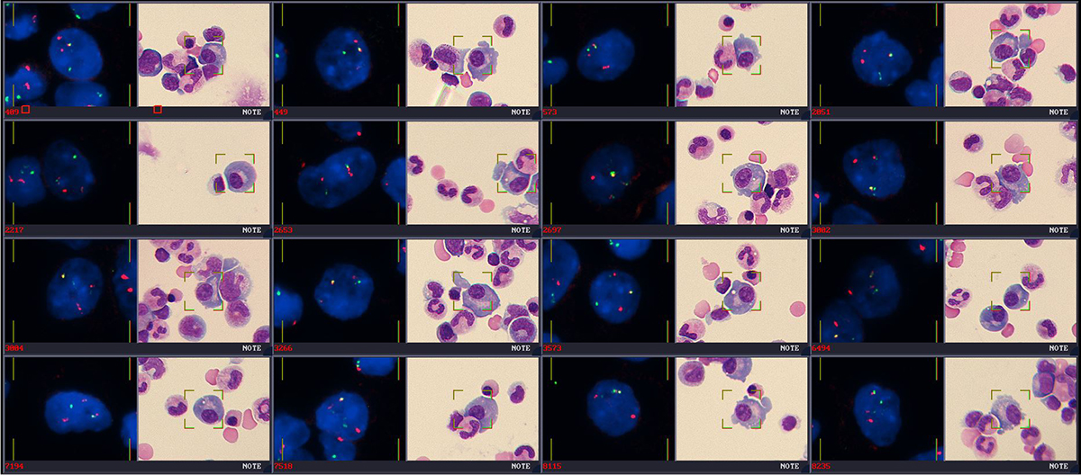
Transparency of this kind also makes disagreement useful. When a reviewer overrides a proposal, the reason is usually visible in the overlay: poor separation, weak signal, an artefact, which is exactly the information a laboratory needs when monitoring a tool over time.
An assistive layer
Automated feature analysis is best understood as an assistive layer over an existing method. It prepares, measures and organises; it does not carry clinical context or professional responsibility. The value comes from pairing consistent measurement with expert judgement, under the laboratory's validation and review procedures.