

Microscope Magnification vs. Optical Resolution: What Matters for Automated Cell Imaging
Higher magnification makes an image bigger. It does not necessarily make it more detailed. The distinction matters when images are acquired for automated analysis.
Magnification is the most visible specification on a microscope, so it is often treated as shorthand for image quality. In automated imaging, where the captured image, not the eyepiece view, is the working material, it helps to separate two properties that behave very differently.
Two different things
Magnification is how much larger an object appears than its physical size. Optical resolution is the smallest separation at which two distinct points can still be distinguished as two. Magnification scales what is already in the image; resolution determines what was captured in the first place.
The practical consequence is that enlarging an image beyond the resolution of the optical system reveals no additional detail. The structures simply become larger and softer, the familiar situation described as empty magnification.
What actually sets resolution
Two factors dominate. The first is the wavelength of light used to form the image: shorter wavelengths can resolve finer detail than longer ones, which is one reason different fluorescence channels do not all behave identically.
The second is the numerical aperture of the objective, which describes the range of angles over which it can collect light. A higher numerical aperture gathers more of the light diffracted by the specimen and therefore resolves finer structure. This is why two objectives with the same nominal magnification can produce noticeably different images, and why immersion objectives exist.
Why this matters for cellular and chromosomal detail
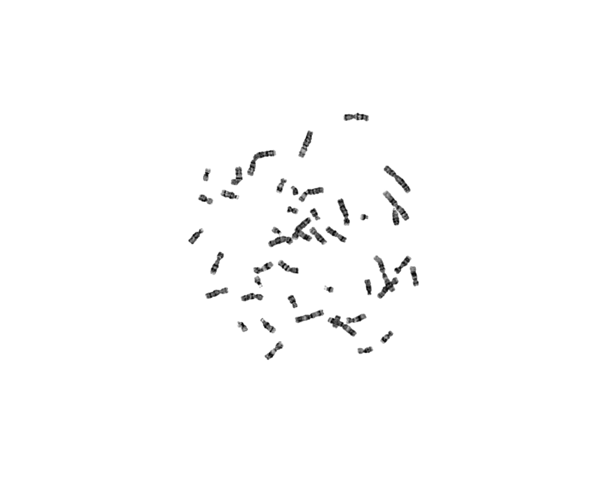
Much of the detail cytogenetics and pathology depend on sits close to the limits of light microscopy: banding patterns along a chromosome, closely spaced fluorescent signals, fine chromatin texture. Whether those features are separable is decided by the optics and acquisition settings, not by how large the image is displayed.
For automated analysis the stakes are higher than for visual review, because measurements are derived from the pixels. If two adjacent signals are not resolved at capture, no downstream algorithm can reliably count them as two. Detection, segmentation and classification all inherit the quality of the acquired image.
Practical points in an automated workflow
- Choose the objective for the smallest structure that must be resolved, considering numerical aperture alongside magnification.
- Keep focus reliable across the scanned area; out-of-focus capture cannot be corrected afterwards.
- Set exposure per channel so that signals are well within range, neither buried in noise nor saturated.
- Sample finely enough that resolved detail is actually recorded in the pixel grid, rather than averaged away.
- Keep acquisition settings consistent between cases so that images, and any measurements taken from them, remain comparable.
- Check illumination uniformity and background periodically as part of routine quality control.
- Review a representative captured image at native scale before committing a full scan configuration.
None of this is exotic; it is ordinary microscopy discipline. But when images feed an automated pipeline, the acquisition step is where most of the achievable quality is decided, and the one step that later processing cannot recover.