
Remote Karyotype Analysis: Building a Connected Review and Collaboration Workflow
Once karyotype images are digital, analysis no longer has to happen beside the microscope. What a connected review workflow looks like, and what laboratories need to plan for.
Karyotype analysis has traditionally been tied to a physical location: the slide, the microscope, and the person sitting at it. Digitisation loosens that constraint. Once metaphase images exist as data, the review step can happen wherever the laboratory permits and by whoever the laboratory authorises.
That shift is organisational as much as technical. The analytical method does not change; what changes is how a case moves between people.
From physical slides to digital cases
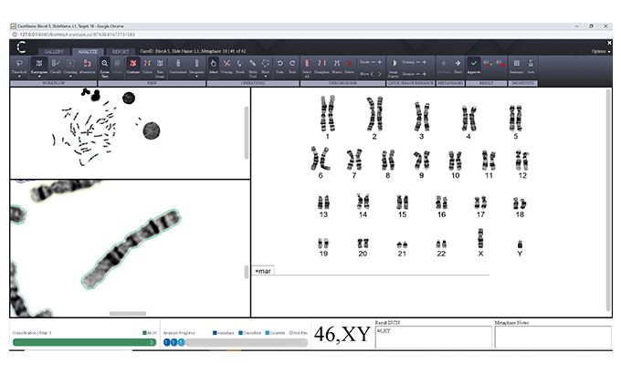
In a manual workflow, the unit of work is the slide. It is scanned at the microscope, analysed by whoever has access to that microscope, and physically transported if a second opinion is needed elsewhere. In a digital workflow the unit of work becomes the case record: a set of captured images with associated metadata, status and history.
What a connected workflow looks like
A connected karyotype workflow generally moves through a recognisable sequence of steps, each of which is visible to the people responsible for it.
- Image capture or import: metaphase images acquired by a scanner or imported into the case.
- Case organisation: images grouped under a case with the identifiers and metadata the laboratory requires.
- Assignment: the case is allocated to an analyst, with workload distributed across the available team.
- Analysis: chromosomes are arranged and edited on screen, with the work saved against the case.
- Expert consultation: a colleague at another site can open the same images without shipping slides.
- Review: a designated reviewer examines the analysis and the images it was based on.
- Reporting: the confirmed result is exported into the laboratory's reporting process, with selected images retained as part of the record.
Operational possibilities
The benefits depend heavily on how a laboratory organises itself, and they are best treated as possibilities to evaluate rather than guarantees.
- Access to expertise that is not physically present at the site holding the sample.
- More flexible distribution of review work across a team or across sites.
- Continuity of review during staffing constraints, absence or peak periods.
- Structured case status, so it is clear what is awaiting analysis, review or reporting.
- Collaboration and teaching on identical case material, including competency assessment.
What a connected workflow does not do is change the amount of analytical judgement a case requires, or the quality of the underlying preparation. Culture, harvest and staining still determine what is available to analyse.
What to plan for
- Permissions: who may open, edit, review and sign out which cases, and how that is enforced.
- Security and data protection: handling of patient-identifiable information in line with applicable requirements.
- Connectivity: sufficient and reliable bandwidth for image review at the sites that need it.
- Standard operating procedures covering remote analysis, second review and escalation.
- Local validation before routine use, on the laboratory's own material.
- Clear responsibility for the final review and result, regardless of where the analysis was performed.
Approached deliberately, remote karyotype analysis is less a new method than a new geography for an existing one — with the accountability structure defined explicitly rather than assumed from physical presence.