

Spectral Bleed-Through in Multi-Color FISH: Causes, Effects and Imaging Considerations
When fluorophore spectra overlap, light from one probe can appear in another channel. What causes bleed-through, how it can affect interpretation, and the imaging factors that reduce it.
Multi-color FISH depends on being able to attribute each fluorescent signal to the correct probe. Spectral bleed-through also described as crosstalk, is the situation where light emitted by one fluorophore is recorded in a channel intended for another. It is a normal optical phenomenon rather than a failure, and recognising it is part of reading multi-color images carefully.
This article is general imaging education. Assay-specific decisions, scoring criteria and acceptance limits should follow each laboratory's own validated procedures.
Why spectra overlap
Fluorophores do not absorb and emit at single wavelengths. Each has a broad excitation spectrum and a broad emission spectrum, and in a multi-color panel those curves are neighbours rather than strangers. Two consequences follow.
- Excitation crosstalk: light intended to excite one fluorophore also partially excites another whose excitation spectrum extends into the same region.
- Emission bleed-through: the long tail of one fluorophore's emission falls inside the detection band of a neighbouring channel.
The closer two fluorophores sit spectrally, and the larger the difference in their signal brightness, the more noticeable the effect tends to be.
How it can affect interpretation
Bleed-through does not invent structures, but it can change how signals appear when channels are viewed as a merged image.
- A strong signal in one channel may produce a faint counterpart at the same position in another, which can read as apparent co-localisation or proximity.
- Channel contamination can raise apparent background and make weak genuine signals harder to distinguish.
- Confidence in separating closely spaced signals of different colours is reduced when part of the intensity may originate from the neighbouring channel.
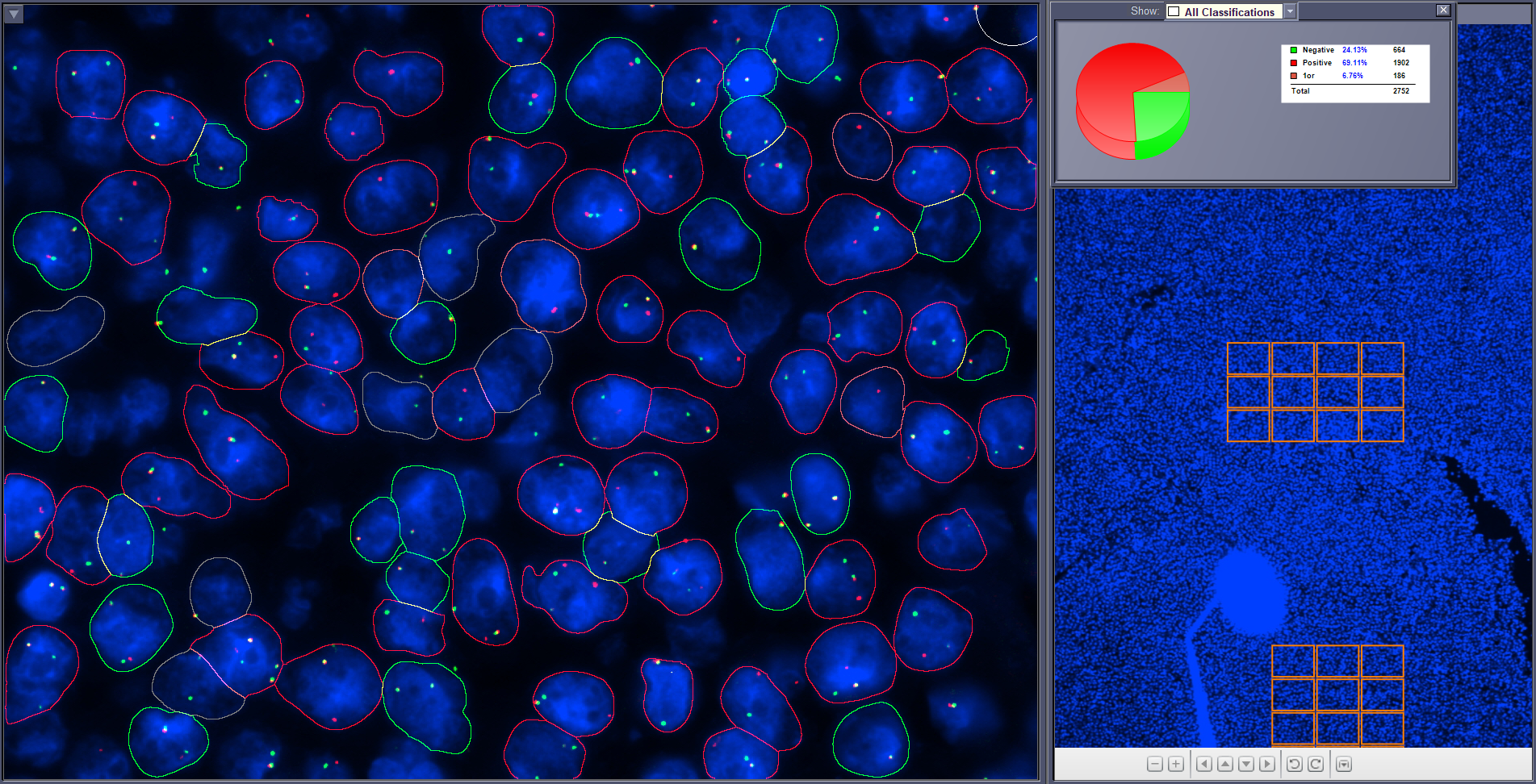
Imaging considerations that help
Most of the practical control over bleed-through sits at acquisition and review rather than in post-processing.
- Filter selection: excitation and emission filter sets chosen for the specific fluorophore combination in use, rather than generic bands.
- Channel-specific acquisition: capturing channels sequentially with settings appropriate to each, instead of a single compromise setting.
- Controlled exposure: avoiding saturation, which exaggerates the spectral tails that cause bleed-through in the first place.
- Appropriate controls: single-fluorophore and unstained preparations make it possible to see what a channel records in the absence of the probe it is meant to detect.
- Review of raw channels: examining individual channels, not only the merged overlay, before concluding that two signals coincide.
- Consistency: keeping acquisition parameters stable so that images and any automated measurements remain comparable across a run.
Where automated analysis fits
Automated systems acquire and present the same optical information, so the same considerations apply. Their contribution is consistency and reviewability: settings are applied uniformly across a slide, and per-channel images remain available so that an analyst can verify any signal that looks equivocal. The judgement about whether a signal is genuine stays with the reviewer.